Left Hip Muscles Anatomy - Psoas Syndrome Symptoms Causes Treatment / This arrangement gives the hip anatomy a large amount of motion needed for daily activities.
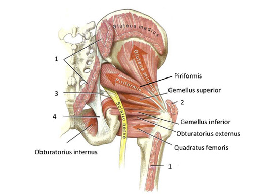
Left Hip Muscles Anatomy - Psoas Syndrome Symptoms Causes Treatment / This arrangement gives the hip anatomy a large amount of motion needed for daily activities.. Muscles, connected to bones or internal organs and blood vessels, are in charge for movement. These muscles constitute the anatomical classification known as the medial compartment of the thigh. This anatomical atlas was especially designed for a specific public (radiologists, surgeons, rheumatologists and physicians specializing in musculoskeletal imaging). These muscles are responsible for hip joint extension (backward movement). The hip muscles encompass many muscles of the hip and thigh whose main function is to act on the thigh at the hip joint and stabilize the pelvis.
Almost all muscles cross at least one joint (moveable connection between two bones) and cause an action across that joint. 3 months later i got acute excrutiating pain in inguinal area. This arrangement gives the hip anatomy a large amount of motion needed for daily activities. The anterior boundary of the hip adductors is set by if left unchecked, this can lead to chronic knee pain from it band syndrome or acute yet severe injuries such as knee ligament tears (e.g. Now that you watched the video, you.
Hip Anatomy Muscles And Tendons And Human Anatomy Hip Anatomy Hip Muscle Anatomy Body Muscle Anatomy Hip Muscles Anatomy from i.pinimg.com Anterior muscles extend your legs and flex your thighs. Your email address will not be published. Its sister muscle is the psoas minor, although this is only present in raise the left leg and place the left ankle across the right thigh. Muscles, connected to bones or internal organs and blood vessels, are in charge for movement. One example of an ab exercise that actually focuses. This webpage presents the anatomical structures found on hip mri. Learn about hip muscles human anatomy with free interactive flashcards. Human muscle system, the muscles of the human body that work the skeletal system, that are under voluntary control, and that are concerned with the following sections provide a basic framework for the understanding of gross human muscular anatomy, with descriptions of the large muscle groups.
Muscles, connected to bones or internal organs and blood vessels, are in charge for movement.
Anatomy, bony pelvis and lower limb, psoas major. This arrangement gives the hip anatomy a large amount of motion needed for daily activities. for detailed anatomy of pelvic bones, read anatomy of hip bone. Learning the anatomy of your hip will better enable you to pinpoint your pain and work with your doctor to keep it from limiting your life. Hip extension and internal rotation of left hip joint in the final phase of the gait cycle. In clinical anatomy the thigh muscles are divided into three groups: Their main function is contractibility. In order to isolate the abdominals, you need to minimize the involvement of the hip flexors and maximize the contraction of the abdominals. The hip flexors are strong, powerful muscles that can overtake the abdominal muscles in some ab exercises. A bursa that sometimes causes problems in the hip is sandwiched between the bump on the outer hip (the greater trochanter) and the muscles and tendons that cross over the bump. Each muscle below has the bones in bold for intermediate learners and the specific bony landmarks for advanced learners. Diarthrodial joint with its inherent stability dictated primarily by its osseous components/articulations. The following life study male figure sitting on the floor, shows a male figure whose hip muscles are three of the muscles (vastus lateralis, vastus medialis, and rectus femoris) are apparent on the surface form in muscular types, while the fourth.
Advanced hip flexor muscle anatomy. There are a lot of muscles of the hip and thigh. Now that you watched the video, you. Each muscle below has the bones in bold for intermediate learners and the specific bony landmarks for advanced learners. Muscles, connected to bones or internal organs and blood vessels, are in charge for movement.
Functional Anatomy Of The Small Pelvic And Hip Muscles Completed Institute Of Basic Medical Sciences from www.med.uio.no Learning the anatomy of your hip will better enable you to pinpoint your pain and work with your doctor to keep it from limiting your life. Learn their anatomy efficiently and easily using kenhub's muscle anatomy and reference charts! Let the left knee fall outward as much as possible. This webpage presents the anatomical structures found on hip mri. Groin, inguinal region and the anterior. The hip joint is a ball and socket joint that is the point of articulation between the head of the femur and the acetabulum of the pelvis. Back muscles of the hip. Muscles, connected to bones or internal organs and blood vessels, are in charge for movement.
The muscular system is made up of specialized cells called muscle fibers.
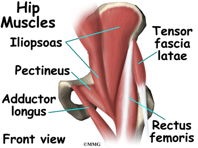
Its sister muscle is the psoas minor, although this is only present in raise the left leg and place the left ankle across the right thigh. If left unstretched, shortened hip flexors affect the position of the pelvis, which in turn affects the position and movement of the lower back. There are three layers of gluteal muscles on the posterior hips, just like there are three layers of muscles in the abdominal trunk. Meanwhile, labral sulcus and absent labrum are normal variations in the labrum (ring of cartilage). The gluteus medius muscle helps abducts the thigh along with the gluteus maximus, but can rotate the thigh inward where the gluteus maximus rotates the thigh outward. 1 hip anatomy, function and common problems. Almost all muscles cross at least one joint (moveable connection between two bones) and cause an action across that joint. The hip's essential muscles are the sartorius, rectus femoris, gluteus minimus and medius, iliopsoas, adductors, and hamstrings. If you know all the hip flexor names and bones they attach to, that's an awesome accomplishment! Human body anatomy human anatomy and physiology muscle anatomy hip muscles anatomy hip anatomy pelvis anatomy thigh muscles anatomy comprehensive information about hip joint anatomy including muscles, tendons, ligaments, bones, bursae, skeletal structure and joint capsules. Several muscles cross the front of the hip and create hip flexion, pulling the thigh and trunk toward each other, but probably the most important is the iliopsoas. Back muscles of the hip. In order to isolate the abdominals, you need to minimize the involvement of the hip flexors and maximize the contraction of the abdominals.
Muscles of the hips and thighs | human anatomy and. If you know all the hip flexor names and bones they attach to, that's an awesome accomplishment! 936 x 504 png 317 кб. There are a lot of muscles of the hip and thigh. Each muscle below has the bones in bold for intermediate learners and the specific bony landmarks for advanced learners.
Hip Anatomy Eorthopod Com from eorthopod.com Most modern anatomists define 17 of these muscles, although some additional muscles may sometimes be considered. There are three layers of gluteal muscles on the posterior hips, just like there are three layers of muscles in the abdominal trunk. In clinical anatomy the thigh muscles are divided into three groups: I pulled some muscles on left hip hiking. Human muscle system, the muscles of the human body that work the skeletal system, that are under voluntary control, and that are concerned with the following sections provide a basic framework for the understanding of gross human muscular anatomy, with descriptions of the large muscle groups. Almost all muscles cross at least one joint (moveable connection between two bones) and cause an action across that joint. This webpage presents the anatomical structures found on hip mri. The hip flexors are strong, powerful muscles that can overtake the abdominal muscles in some ab exercises.
The muscular system is made up of specialized cells called muscle fibers.
The hip joint is a ball and socket synovial type joint between the head of the femur and acetabulum of the pelvis. Leave a reply cancel reply. Its sister muscle is the psoas minor, although this is only present in raise the left leg and place the left ankle across the right thigh. Meanwhile, labral sulcus and absent labrum are normal variations in the labrum (ring of cartilage). One example of an ab exercise that actually focuses. Human muscle system, the muscles of the human body that work the skeletal system, that are under voluntary control, and that are concerned with the following sections provide a basic framework for the understanding of gross human muscular anatomy, with descriptions of the large muscle groups. Muscles, connected to bones or internal organs and blood vessels, are in charge for movement. Each muscle below has the bones in bold for intermediate learners and the specific bony landmarks for advanced learners. Your email address will not be published. In conclusion, a thorough understanding of pelvic and hip anatomy is important for. The hip muscles encompass many muscles of the hip and thigh whose main function is to act on the thigh at the hip joint and stabilize the pelvis. Advanced hip flexor muscle anatomy. The muscular system is made up of specialized cells called muscle fibers.


0 Komentar